| The HAXPEEM (Hard X-ray Photoemission Electron Microscope) instrument currently installed at beamline P22 (PETRA III, DESY, Hamburg) is an energy filtering photoemission microscope of NanoESCA design, developed and funded (BMBF project, grant #03SF0445) in collaboration between FZ Jülich and DESY. In order to cope with the high kinetic energies involved in HAXPES (Hard X-ray Photoemission Spectroscopy), the sample bias as well as the extractor potential has been scaled up to cover a range of 0-10keV kinetic energy. |
| The instrument uses sample holders similar to the Focus PEEM sample holders which accept flat specimen of dimension 10mm x 10mm x 1mm. The storage chamber is equipped with 8 slots, 6 of which are free to use. Quick sample exchange is done using a wobble stick. |
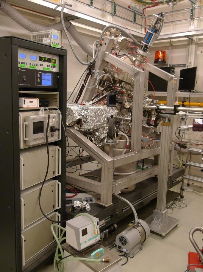 HAXPEEM in measurement position at P22
|
 Load lock and storage chamber
|
|
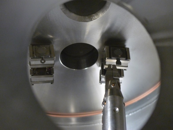 Sample storage
|
Challenges: Photoionization Cross-Sections, Instrument Transmission
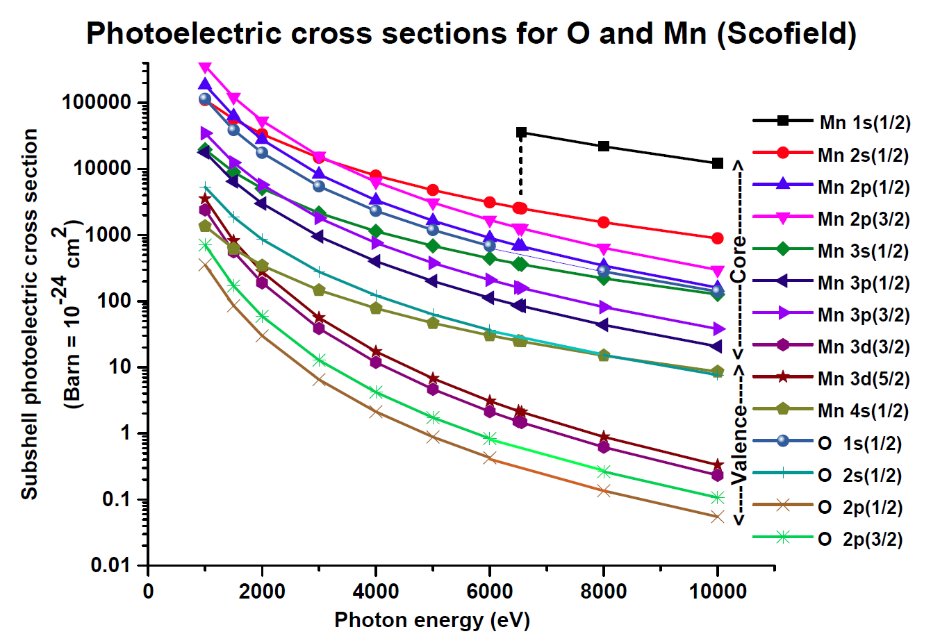
| Taking HAXPES to 2D comes with some challenges. The cross sections for photoemission drop substantially at higher photon energies compared to laboratory UPS or soft x-ray sources. Countering this signal loss with the higher photon flux delivered by the synchrotron beamline also finds a limit, since the number of low-energy electrons increases as well, leading to space charge effects at the sample surface. Additionally, the electron optics transmission function favors low energies. To cope with the resulting low signal strength, the instrument is equipped with a software based single electron counting detection. Moreover, the stability of the instrument and beamline enable long exposition times. |
|
Photoionization cross sections for O and Mn up to 10keV photon energy1 |
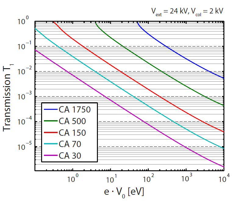
Calculated transmission of the objective lens vs electron kinetic energy2 |
Benefits: 2D spatial resolved electron spectroscopy from below the surface
| As a full-field imaging system, the HAXPEEM captures energy filtered images of the complete field of view at once. Below are some examples taken from recent projects. |
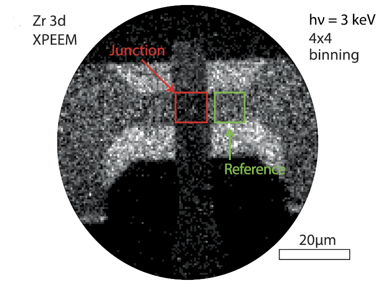
HAXPEEM of a zirconiumoxide resistive switching device imaged on Zr 3d. Bottom and top electrode of the crossbar design overlap in the region marked in red (junction) in the image center3 |
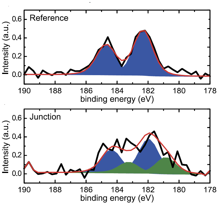
Zr 3d spectra extracted from selected areas in an image series in the FoV shown on the left. |
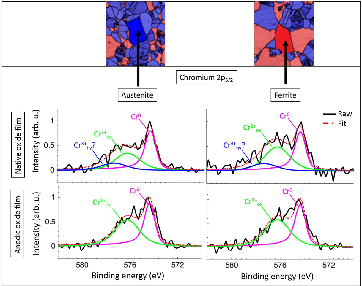
HAXPEEM Cr 2p spectra from different micron-sized grains of a duplex steel alloy. Top row: colorized EBSD maps identifying Austenitic and Ferritic phases. Middle row: Cr 2p spectra of native oxide. Bottom row: Cr 2p spectra after anodic polarization in NaCl.4 |
|
References:
- P. H. Scofield, Lawrence Livermore Laboratory Report UCRL-51326 (1973)
- M. Patt, C. Wiemann, N. Weber et al., Review of Scientific Instruments 85, 113704 (2014)
- A. Kindsmüller, C. Schmitz, C. Wiemann et al., APL Materials 6, 046106 (2018)
- M. Långberg, C. Örnek, F. Zhang et al., Journal of The Electrochemical Society, 166 (11) C3336-C3340 (2019)