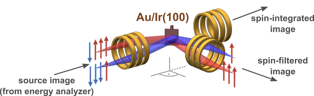
The promise of the spin-resolved experiments throughout the surface Brillouin zone has been severely hindered for decades, due to the extremely low spin-detection efficiency [1]. Major advances have been made via the recent invention of the multi-channel spin-polarization detector, the so-called imaging spin filter [2]. It provides a stunning increase in spin-detection efficiency by four orders of magnitude, in comparison to conventional single-channel electron spin detectors [1,2].
References
|
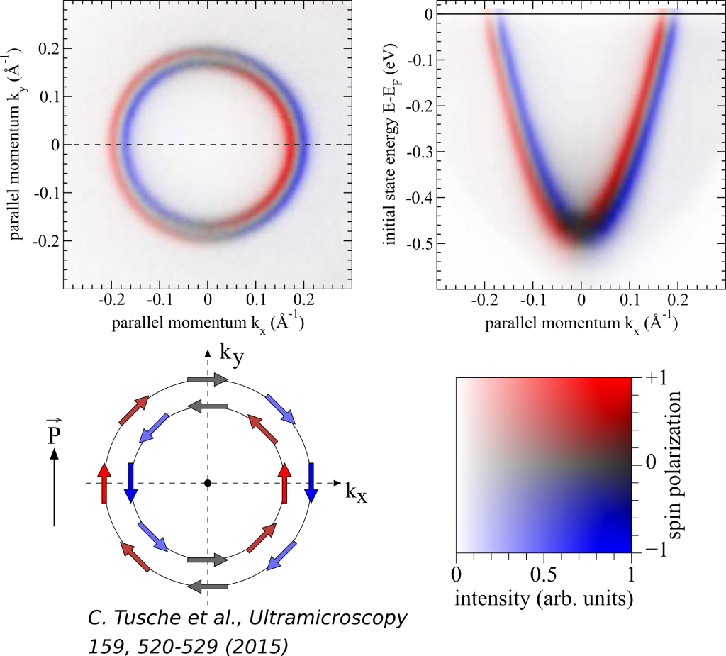
| Measured spin polarization and intensity map at EF of the Au(111) Rashba-split surface state excited by p-polarized 6.05 eV photons with respect to the quantization axis indicated by P. Note the 2D color code for intensity and spin polarization. |
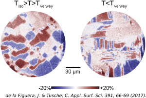
| For the first time, on magnetite (001) the near surface real-space reorganization of the magnetic domains has been followed in detail by utilizing spin-resolving momentum microscopy, both upon crossing the Verwey transition when cooling and when heating. The illumination is performed by a He I lamp, and the energy of the secondary electrons is Ef + 5.82 eV. |