 |
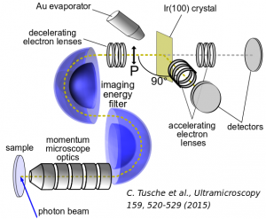
The working principle of our spin resolving momentum microscope system, consisting of He-cooled sample stage, imaging electron optics, two hemispherical analyzers, and detection branches for spin-integral and spin-filtered imaging. The Ir(100) crystal can be inserted/retracted after the 2nd HDA. |
| Based on the electron optical properties of the momentum microscope optics, electrons emitted from the sample surface in all directions are simultaneously collected, and imaged on the 2D detector. These “momentum images” are always scaled linearly in the kx and ky coordinates, and represent a direct view of the reciprocal space, for instance, as a section through the Fermi surface of the sample. |
Bandstructure imaging from overview to highest resolution
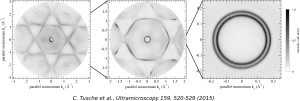
| Photoemission pattern from the Au(111) surface at the Fermi energy from full image of the surface Brillouin zone to the detail of the Rashba-split Shockley surface state, using a higher momentum magnification with the energy resolution down to 12 meV and a momentum resolution of 5×10⁻³ Å−1. Note that a momentum-image covering the diameter of 2 Å-1 can be collected from an area as small as 1 μm. |
High resolution energy filter
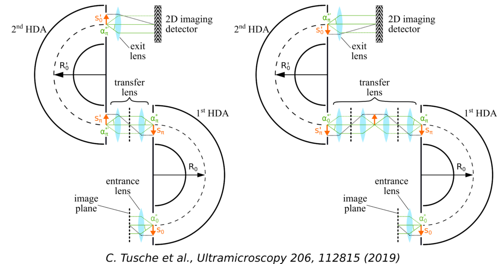
| Newly developed momentum microscopy in our group has realized a switchable non-inversion transfer lens in a so-called double pass configuration (right), situated between two electrostatic hemispherical deflection analyzer (HDA). That has accomplished a four times increased transmission at comparable energy and momentum image resolution compared to previous approaches (left). |